31P-MRS membrane permeability markers relate to disease activity in BMD
01 Dec 2025
I just published a new article as last author in NMR in Biomedicine Skeletal muscle membrane permeability markers derived from 31P-MRS may reflect disease activity in Becker muscular dystrophy. This was a close collaboration between me and my former colleagues at Leiden University Medical Center, where we show the potential of phosphorus-31 magnetic resonance spectroscopy for capturing early muscle changes in neuromuscular disorders.
A brief abstract is given below:
We compared several candidate biomarkers for disease activity between patients with Becker muscular dystrophy and controls: intracellular ionised magnesium ([Mg 2+]), phosphodiesters (PDE), and
weighted pH measures from phosphorus-(31P)-MRS; and membrane permeability derived from the random permeable barrier
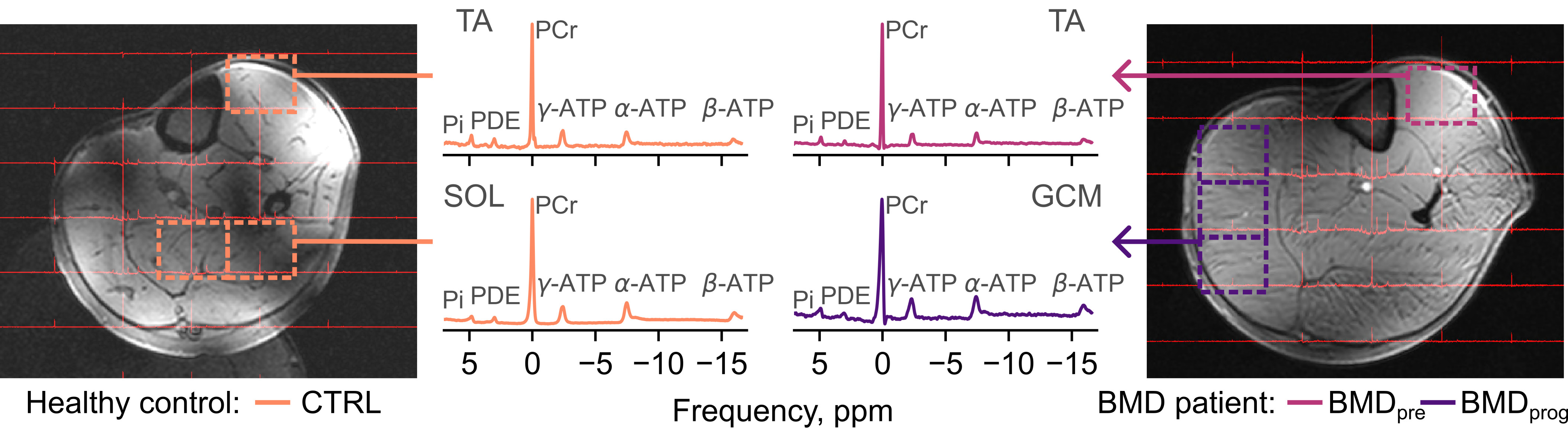
model (RBPM), as applied to diffusion-tensor-(DT)-MRI data. We performed 7 tesla 31P-MRS and 3 tesla DT-MRI in the left lower leg of
23 participants with BMD and 14 healthy controls, estimating [Mg2+], PDE/γ-ATP, weighted pH and RPBM permeability. Follow-up scans at 24 months were performed in a
subset of participants. Muscles in patients with BMD were regarded as likely to be ‘preserved’, with fat fractions
≤ 13.5% and as ‘progressing’, with fat fractions between 13.5% and 81.5%. Muscles with fat fractions ≥ 81.5% were excluded from
further analyses. We observed decreased [Mg2+] in BMD pre and BMD prog compared to healthy controls, whereas PDE/γ-ATP
and weighted pH were increased in these muscles. RBPM-measured permeability did not differ between groups. In patients with BMD, 31P-MRS demonstrates re-
duced [Mg2+] and increased weighted pH in the lower leg muscles versus controls, suggesting greater membrane permeability—a
potential disease activity biomarker independent of disease phase. PDE/γ-ATP was also significantly increased in progressing
and preserved muscle. Incorporating 31P-MRS in therapeutic trials will help to further establish its use as a response biomarker.