01 Dec 2025
I just published a new article as last author in NMR in Biomedicine Skeletal muscle membrane permeability markers derived from 31P-MRS may reflect disease activity in Becker muscular dystrophy. This was a close collaboration between me and my former colleagues at Leiden University Medical Center, where we show the potential of phosphorus-31 magnetic resonance spectroscopy for capturing early muscle changes in neuromuscular disorders.
A brief abstract is given below:
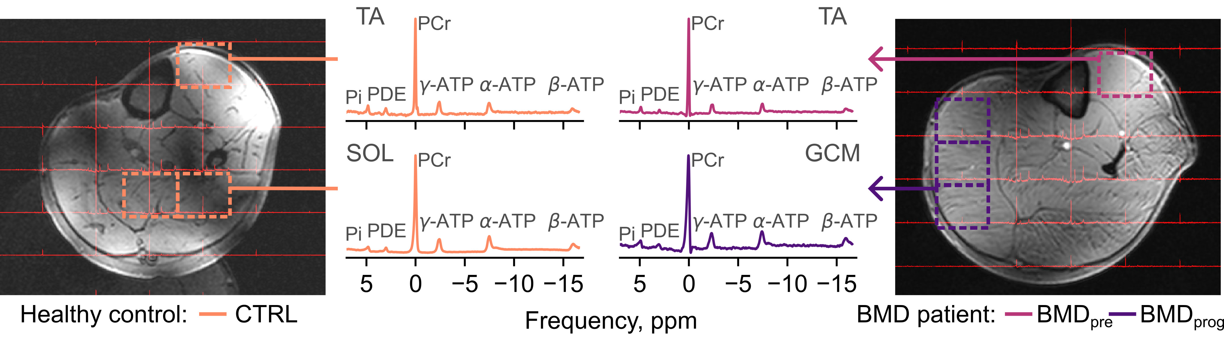
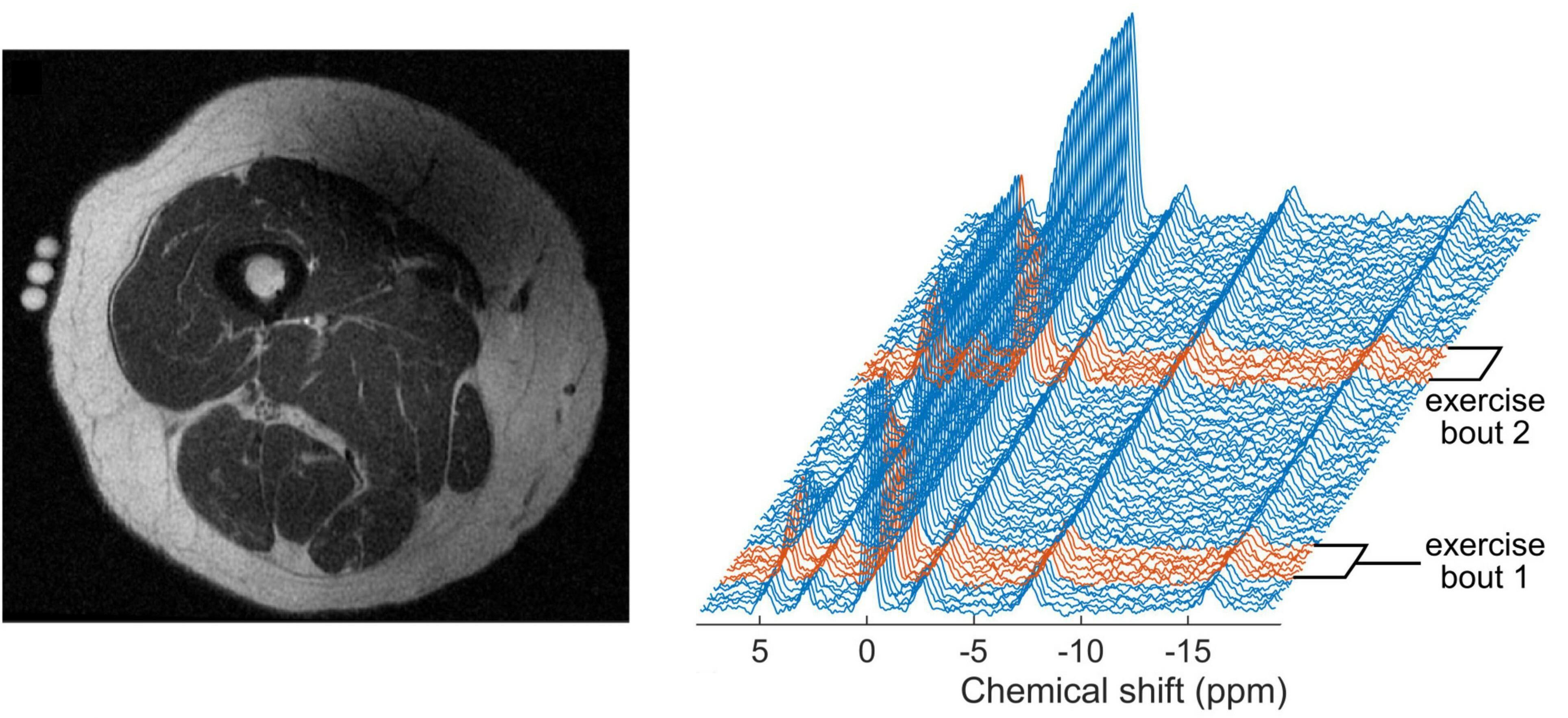
We compared several candidate biomarkers for disease activity between patients with Becker muscular dystrophy and controls: intracellular ionised magnesium ([Mg 2+]), phosphodiesters (PDE), and
weighted pH measures from phosphorus-(31P)-MRS; and membrane permeability derived from the random permeable barrier
model (RBPM), as applied to diffusion-tensor-(DT)-MRI data. We performed 7 tesla 31P-MRS and 3 tesla DT-MRI in the left lower leg of
23 participants with BMD and 14 healthy controls, estimating [Mg2+], PDE/γ-ATP, weighted pH and RPBM permeability. Follow-up scans at 24 months were performed in a
subset of participants. Muscles in patients with BMD were regarded as likely to be ‘preserved’, with fat fractions
≤ 13.5% and as ‘progressing’, with fat fractions between 13.5% and 81.5%. Muscles with fat fractions ≥ 81.5% were excluded from
further analyses. We observed decreased [Mg2+] in BMD pre and BMD prog compared to healthy controls, whereas PDE/γ-ATP
and weighted pH were increased in these muscles. RBPM-measured permeability did not differ between groups. In patients with BMD, 31P-MRS demonstrates re-
duced [Mg2+] and increased weighted pH in the lower leg muscles versus controls, suggesting greater membrane permeability—a
potential disease activity biomarker independent of disease phase. PDE/γ-ATP was also significantly increased in progressing
and preserved muscle. Incorporating 31P-MRS in therapeutic trials will help to further establish its use as a response biomarker.
13 May 2023
My latest article, Age-related changes in human skeletal muscle microstructure and architecture assessed by diffusion-tensor magnetic resonance imaging and their association with muscle strength - published in Aging cell - was a labour of love that began back in 2015 as part of the GESTALT study of ageing. We show that diffusion-MRI-measured skeletal muscle microstructure (fractional anisotropy and mean diffusivity) and architecture (pennation angle, fascicle length, curvature, and PCSA) are associated with age and with muscle strength. This will prove important for understanding the early stages of sarcopenia - the loss of muscle mass and strength with ageing.
A bried summary of the paper is given below:
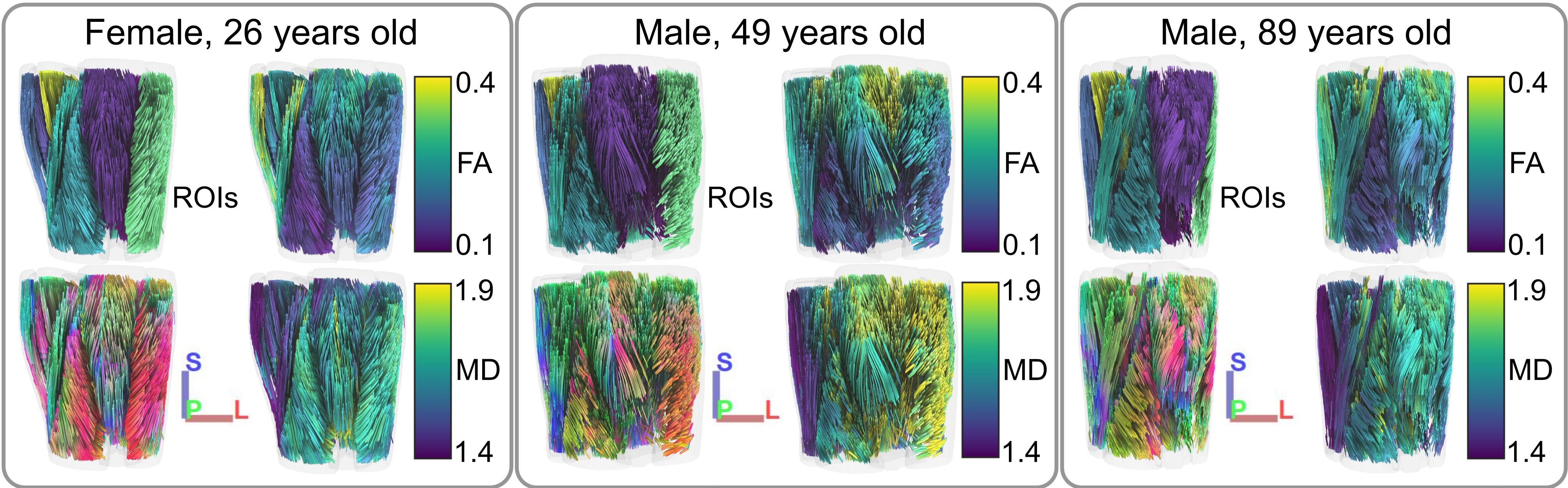
We used diffusion-tensor MRI (DT-MRI) to probe muscle microstructure and architecture in a large healthy-ageing cohort, with the aim of characterizing age-related
differences and comparing to muscle strength. We recruited 94 participants (age range = 22–89 years) and measured
microstructure parameters—fractional anisotropy (FA) and mean diffusivity (MD)—in 12 thigh muscles, and architecture parameters—pennation angle,
fascicle length, fiber curvature, and physiological cross-sectional area (PCSA)—in the rectus femoris (RF) and biceps femoris longus (BFL).
Knee extension and flexion torques were also measured for comparison to architecture measures. FA and MD were associated with age, as were pennation angle, fiber curvature,
fascicle length, and PCSA in the RF, while in the BFL only curvature and fascicle length were associated with age. In the RF, pennation angle and PCSA were
associated with strength; in the BFL, only PCSA was associated with strength. Our results show skeletal muscle architectural changes with ageing
and intermuscular differences in microstructure. DT-MRI may prove useful for elucidating muscle changes in the early stages of sarcopenia and
monitoring interventions aimed at preventing age-associated changes in muscle that lead to functional impairment.
03 May 2023
My recent work - Diffusion-tensor magnetic resonance imaging captures increased skeletal muscle fibre diameters in Becker muscular dystrophy - shows that diffusion MRI is able to capture the increased muscle fibre sizes arising from repeated cycles of damage and repair in Becker muscular dystrophy (BMD). Such measurements are normally only accessible via muscle biopsies, so this non-invasive approach has important implications for monitoring of new treatments for BMD and other neuromuscular diseases.
Here’s the abstract:
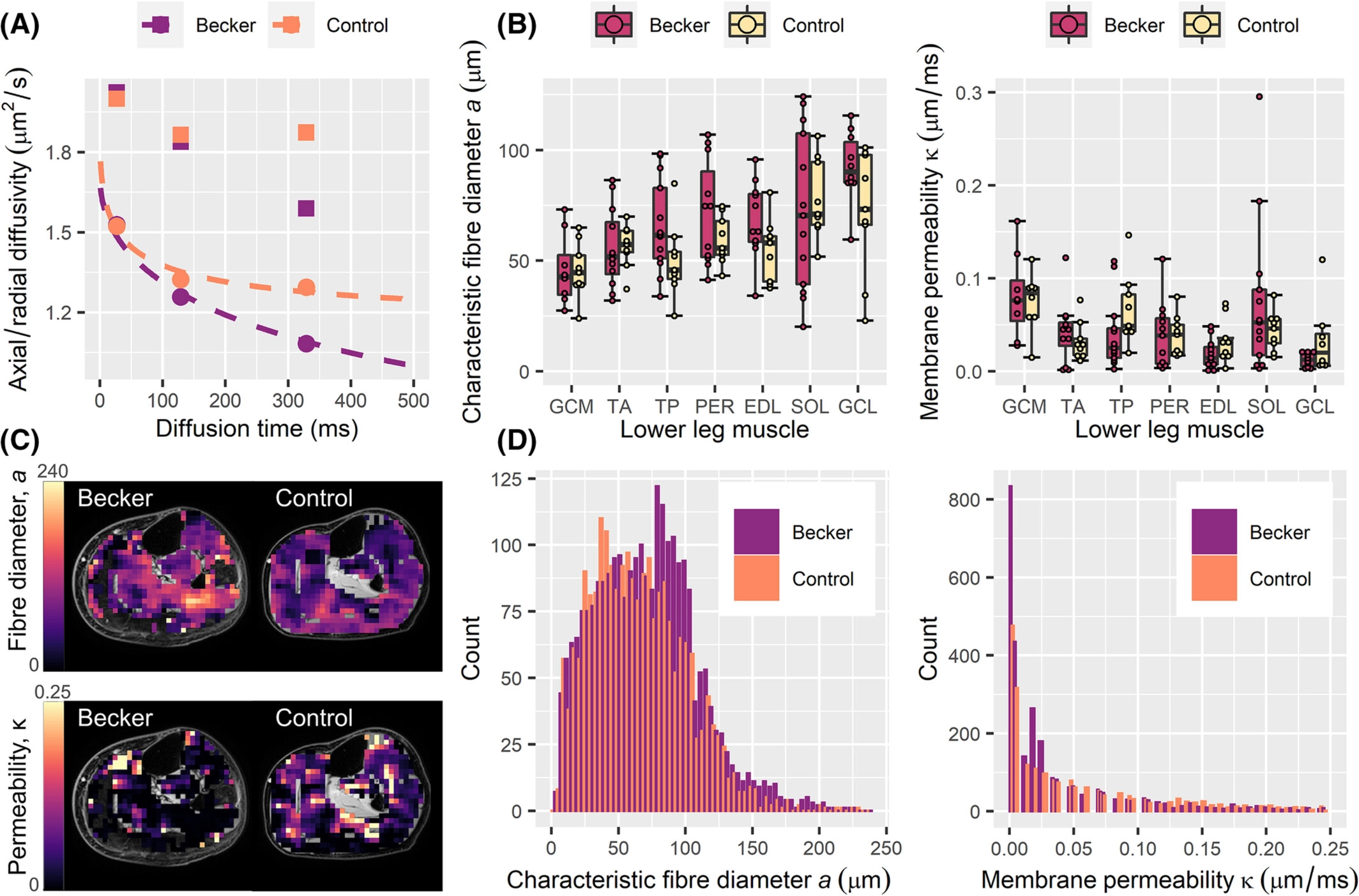
Becker muscular dystrophy (BMD) is characterized by slow, progressive muscle damage and muscle weakness. Hallmarks include fibre-size variation and replacement of muscle with fibrous and adipose tissues, after repeated cycles of regeneration. Muscle histology can detect these features, but requires biopsies. Diffusion-tensor magnetic resonance imaging (DT-MRI) is a potential non-invasive alternative that can calculate muscle fibre diameters when applied with the random permeable barrier model (RPBM). We included 13 BMD patients and 9 controls, who underwent DT-MRI of in the lower leg. Tibialis anterior muscle biopsies were taken in 38 BMD patients and 15 controls. Laminin and Sirius-red stainings were performed to evaluate muscle fibre morphology and fibrosis.
RPBM fibre diameter was larger in BMD patients. Laminin staining agreed with the RPBM, showing larger median fibre diameters in patients than in controls. Patients also showed more intra-individual fibre diameter variability than controls and larger fibrosis areas.
DT-MRI can quantify changes in muscle fibre size that are associated with regeneration without the need for biopsies - showing promise as an imaging biomarker for muscular dystrophies.
31 Jan 2022
Our new article, titled Evaluation of Acute Supplementation With the Ketone Ester deltaG in Healthy Volunteers by Cardiac and Skeletal Muscle 31P Magnetic Resonance Spectroscopy, has just been published in Frontiers in Physiology as part of the Research Topic “New Views on Old Molecules: MR Spectroscopic Techniques & Insights on Physiology”. Here’s a brief synopsis of the research:
In this acute intervention study, we investigated the potential benefit of ketone supplementation in humans by studying cardiac PCr to ATP ratios and skeletal muscle PCr recovery using 31P-MRS before and after ingestion of a ketone ester drink. We recruited 28 healthy adults: 12 age 23–70 for cardiac 31P-MRS, and 16 age 60–75 for muscle 31P-MRS. Baseline and post-intervention 31P-MRS scans were performed in one visit, where 25 g of the ketone monoester, deltaG®, was administered after the baseline scan. Post-intervention 31P-MRS was timed 30 min after deltaG® ingestion. In blood samples, post-intervention glucose, lactate and non-esterified fatty acids decreased (−28.8%, −28.2%, and −49.1%, respectively), while ketone body D-beta-hydroxybutyrate increased from mean (SD) 0.7 (0.3) to 4.0 (1.1) mmol/L. Cardiac PCr/ATP and muscle metabolic parameters did not change. Acute ketone supplementation caused mild ketosis in blood, with drops in glucose, lactate, and free fatty acids; however, these changes were not associated with 31P-MRS measures in heart or muscle. Future work may investigate longer-term ketone supplementation on tissue energetics in groups with compromised mitochondrial function.
20 Sep 2020
We recently published an article, titled Age and Muscle Function Are More Closely Associated With Intracellular Magnesium, as Assessed by 31P Magnetic Resonance Spectroscopy, Than With Serum Magnesium, in Frontiers in Physiology, Striated Muscle Section. Here’s a summary of our work:
Total serum magnesium is a common clinical measurement for assessing magnesium status; however, magnesium in blood represents less than 1% of the body’s total magnesium content. We measured intramuscular ionised magnesium by 31P-MRS and tested the hypothesis that this measure better correlates with muscle function and captures more closely the effect of aging than serum magnesium. Data were collected from 441 participants (age 24–98 years) in the BLSA, a study of healthy aging. We showed that muscle ionised magnesium was negatively associated with age and positively associated with knee-extension strength, while serum magnesium was not associated with either. Muscle magnesium was lower in women than in men, perhaps due to chronic latent magnesium deficiency that is not detected via serum magnesium. Based on these findings, we suggest that intramuscular ionised magnesium from 31P-MRS is a better clinical measure of magnesium status than total serum magnesium; it could be measured when muscle weakness of unidentified aetiology is detected, and to monitor the effectiveness of magnesium interventions, including supplementation.
This work was featured in Mail Online on the 7th of September, 2020.