DT-MRI as a potential biopsy alternative in Becker muscular dystrophy
03 May 2023
My recent work - Diffusion-tensor magnetic resonance imaging captures increased skeletal muscle fibre diameters in Becker muscular dystrophy - shows that diffusion MRI is able to capture the increased muscle fibre sizes arising from repeated cycles of damage and repair in Becker muscular dystrophy (BMD). Such measurements are normally only accessible via muscle biopsies, so this non-invasive approach has important implications for monitoring of new treatments for BMD and other neuromuscular diseases.
Here’s the abstract:
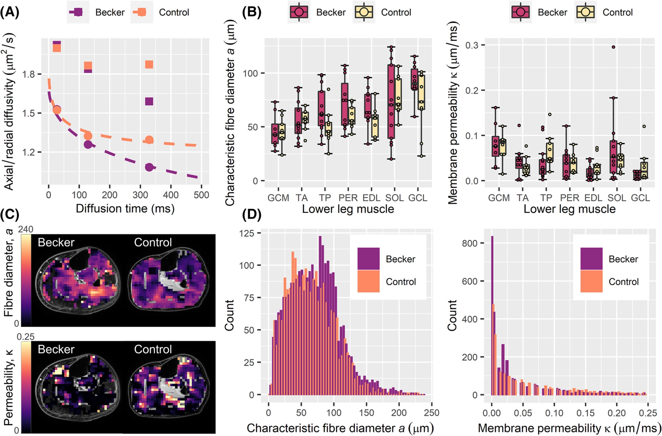
Becker muscular dystrophy (BMD) is characterized by slow, progressive muscle damage and muscle weakness. Hallmarks include fibre-size variation and replacement of muscle with fibrous and adipose tissues, after repeated cycles of regeneration. Muscle histology can detect these features, but requires biopsies. Diffusion-tensor magnetic resonance imaging (DT-MRI) is a potential non-invasive alternative that can calculate muscle fibre diameters when applied with the random permeable barrier model (RPBM). We included 13 BMD patients and 9 controls, who underwent DT-MRI of in the lower leg. Tibialis anterior muscle biopsies were taken in 38 BMD patients and 15 controls. Laminin and Sirius-red stainings were performed to evaluate muscle fibre morphology and fibrosis.
RPBM fibre diameter was larger in BMD patients. Laminin staining agreed with the RPBM, showing larger median fibre diameters in patients than in controls. Patients also showed more intra-individual fibre diameter variability than controls and larger fibrosis areas.
DT-MRI can quantify changes in muscle fibre size that are associated with regeneration without the need for biopsies - showing promise as an imaging biomarker for muscular dystrophies.